
Our research institute is getting its new auditorium. Every time I pass through the construction site, I watch this structure taking its shape, gradually growing each day. It started with an empty plot, a canvas waiting for its masterpiece. Then, bit by bit, the iron and concrete added life into it. The process is meticulous; a little miscalculation and you end up with a staircase leading to a wall instead of a door.
As a brain development researcher, it makes me think about the creation of our own brain. Known as the most complex tissue of our body, the brain is a testament to millions of years of evolution. It is a seat of our consciousness, our intellect, our memory. And its complexity is notably attributed to its structure formed by densely packed inter-connected neurons, enclosed within a limited space inside the skull.
How is this intricate structure formed? Who are the builders and building blocks in this quest for brain development and what happens when things go awry?
The story of brain development begins early in our lives, even before we are born. As embryos, our brains start as a sheet of cells. These cells are our versatile builders called as “neural stem cells”. They have the potential to shape the entire nervous system. These cells can “self-renew”, that is, they can divide and produce more stem cells. This ensures that there are always enough of them available. However, they also have the ability to “differentiate”, that is, they transform into neurons or glia.
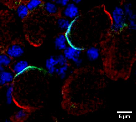
Red-Present in the upper part of cells required for asymmetric division
Green-Present in the lower part of cells helps in asymmetric division
Blue-Present in lower part of stem cells and after division it enters the progenitor cells. It gives cues to the cells to differentiate. PC: Aishwarya Arun, Sonal Nagarkar Jaiswal lab
The stem cells’ dilemma between making their own kind versus making neurons or glia is solved by the asymmetric division of the stem cells. The contents of a neural stem are unequally distributed between its two daughter cells. The bigger cell retains neural stem cell abilities and continues generating more cells. The smaller cell is called as the “progenitor” cell. It gets the molecular cues to differentiate, and it divides itself into neurons or glia.
The neurons transmit information in the form of electrical signals. They have branches called as dendrites that receives the information, and a long tail-like axon that sends the information to other neurons. Using the axons and dendrites, neurons form thousands of connections with other neurons and helps in transmission and processing of information.
On the other hand, the glial cells provide physical support, nutrient supply and insulation to the neurons helping them function smoothly. Some of them also act as the brain’s immune system.
Contrary to earlier beliefs, neurogenesis, the process of birth of neurons, goes on lifelong. The rate does decline with age, which depends on several factors like exercise, environment and diet. In specific brain regions, like the hippocampus known for its role in learning and memory, new neurons continue to be born throughout adulthood. Also, this process helps to repair the brain after injuries.
However, there needs to be a balance between the number of neural stem cells renewing its own population and those forming neurons. For instance, a decline in neural stem cells in brains, results in neurodevelopmental disorders like microcephaly or neuro-psychiatric disorders like epilepsy and autism. On the contrary, if the neural stem cells lose their ability of differentiation, they will end up renewing themselves uncontrollably resulting in malignancies such as brain tumour. This balance between neural stem cell renewal and differentiation is maintained by different genes, their protein products and molecular pathways.
To understand these systematically, scientists need systems that mimic conditions similar to the human brain, and tools to study and manipulate them. One such system are the fruit flies, Drosophila melanogaster, the tiny insects that are hovering over the fruits on your kitchen table.
The neural stem cells of developing fly brain have emerged as a valuable model to study neurogenesis. Many of the genes involved in neurogenesis in flies are also present in humans, and serve similar functions. Scientists can manipulate such genes in fruit fly owing to its simple genetic makeup compared to humans. This gives them insights into the functions of those genes. Along with this, the fly brain is less complex and has well-defined lineages of neurons. All the cells types in a lineage have their distinct protein composition. Scientists can follow the entire lineage from stem cell to neuron by tracking such proteins. In addition to this, it only takes 10 to 12 days for a fly to become an adult. This accelerates the pace of scientific experiments. All of these help brain researchers like me to study neurogenesis.
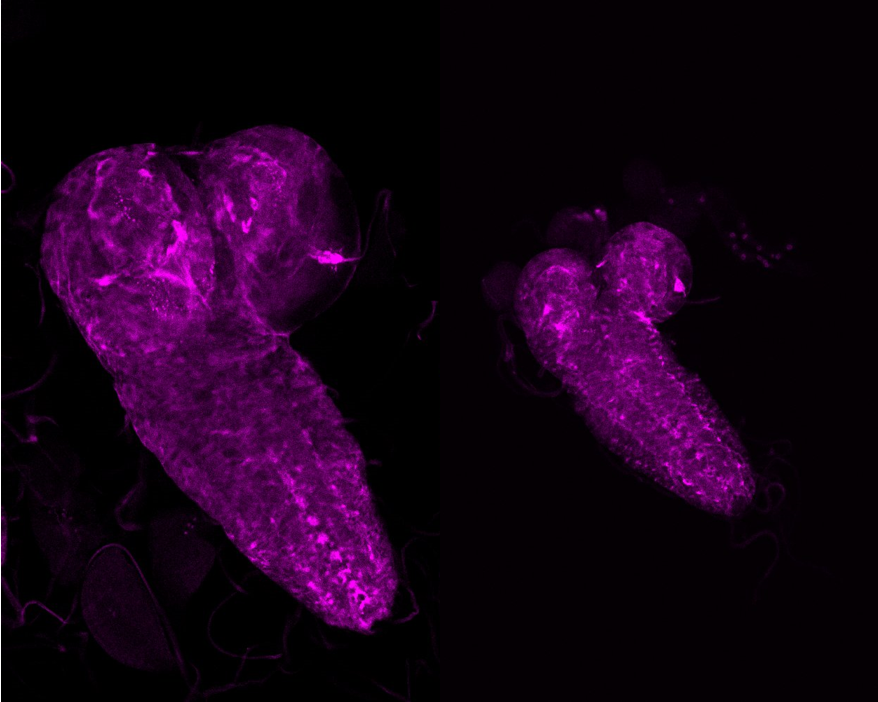
I am currently working on microcephaly or small brain condition. This is a lethal condition where the brain doesn’t grow properly, resulting in a small brain. I have genetically mutated flies to have small brains. The genes that I work with are also present in humans, and their mutations lead to microcephaly too. Some of these mutations affects the neural stem cell size, causes abnormal cell division or even untimely death of stem cells. Using microscopy techniques, I examine any changes in the patterns and levels of proteins required for neural stem cell growth and functioning upon the mutations, and thus, the role of such genes in causing microcephaly condition. I hope such interventions will encourage brain repair and recovery in future times.
Other researchers have also used fruit flies to study intellectual disabilities and behavioural challenges caused by hampered brain development. Scientists can record fly behaviours like sleeping, sensing their environment, responding to smell and visual stimuli. While this may help in drawing parallels with human behavioural impairments, these also offer a model system to test potential drugs for treating brain related disorders.
But, fruit fly and human brains also have their own unique differences. Human brain is much larger than a fly brain, and humans undertake much more complex activities than what we understand of flies. There are differences in the cellular processes too. For example, unlike the asymmetric division of fruit fly neural stem cells, the human cells divide both symmetrically and asymmetrically. Hence, the information obtained from fly model system requires careful consideration.
Nonetheless, flies are already helping scientists and doctors solve real-life challenges. A few years back, a family in the USA had both of their children affected by microcephaly. When they approached hospital authorities, scientists took on the case and identified a mutation in a particular gene through genome sequencing. They turned to a similar gene in fruit flies. They mutated the fly gene, and found it caused microcephaly in fly babies. They confirmed the role of the mutated gene when they could fix fly’s microcephaly condition by introducing a normal human gene. The knowledge also helped the family later on in conceiving a healthy child, without the mutation through IVF.
The tiny fruit flies can be a starting point in cracking the mysteries of brain development. It’s like studying a simple practice puzzle to understand how a big, complex puzzle works to transform into a maestro accessing our thoughts, emotions and co-ordination of various bodily functions. Like as our new auditorium will soon become a fully realized space. All thanks to the remarkable journey of plans, cues and builders.