
In the early 2000s, scientists achieved one of the greatest feats in human history: we sequenced the entire human genome. It felt like we’d finally found the ultimate cheat code for human health. When it came to severe neurological conditions—from intractable epilepsy to complex neurodevelopmental disorders—we could suddenly pinpoint the exact genetic mutation causing the disease.
These genetic mutations are like typos—mistakes that can make or break the functional molecules of life: proteins. We had found the typos. We thought the cures would immediately follow. But a strange, frustrating reality quickly set in: knowing the genetic code of a disease actually tells you almost nothing about how to physically cure it.
Which raises a massive question: If we know exactly which gene is broken, why can’t we just fix the disease?
The Shape-Shifting Machines
Life doesn’t happen in a one-dimensional string of code; it happens in the three-dimensional, physical world. To fix a disease, we have to look past the genetic typo and examine the actual, physical machinery it builds: proteins. And nowhere are these molecular machines more complex—or more devastating when broken—than in the human nervous system.
Take the brain. It contains billions of neurons communicating at lightning speeds—but here’s the twist: neurons don’t actually touch. There are microscopic gaps between every single neuron, called synapses. Electrical signals cannot jump these gaps on their own. Instead, they’re converted into chemical messengers called neurotransmitters, which drift across to the other side. Waiting there are massive, intricate protein structures that form giant channels—ionotropic neurotransmitter receptors—the ultimate gatekeepers of the brain.
These aren’t static doorways. Each receptor is a colossal, tangled cluster of thousands of amino acids, folded into a precise 3D shape. When a messenger binds to it, the entire structure physically contorts—twisting and shifting to open a microscopic pore, allowing charged ions to flood the cell and spark an electrical impulse. All of this happens in a fraction of a millisecond.
So, what does a genetic “typo” actually do to the receptors?
Your DNA is essentially a recipe that tells the cell exactly which amino acids to string together to build the receptor. When a mutation occurs, the cell reads a faulty instruction and accidentally swaps out just one of those thousands of building blocks.
Imagine building a massive, moving Lego engine, but just one single gear is swapped for a slightly bulkier one. The engine might still look fine from a distance—but when you run it, the gears grind and jam. The exact same thing happens to the receptor. That one wrong amino acid means the protein physically cannot fold or move correctly. The bulky block might wedge the pore permanently open, flooding the brain with a lethal surge of electricity. Or it might alter the delicate rhythm of opening and closing so that signals simply die.
You can read the DNA sequence a million times, but it will never tell you exactly how that 3D protein is physically wedged. To design a cure, you have to actually photograph the molecule—at scales where you can see the wrongly-swapped gear. But this is extraordinarily hard, because of just how small proteins are.
The Microscope Problem
Your typical light microscope uses photons. When light shines on an object, it bounces off, passes through glass lenses, and forms an image. But as objects get smaller, you hit a hard, physical limit.
Light can be imagined as waves, and the distance between wave peaks is called the wavelength. Visible light spans roughly 400 to 700 nanometres. The proteins we want to examine are only about 20 nanometres wide—roughly 40 times smaller than the light itself. So instead of bouncing off, light waves simply roll right past the protein. You could build the most perfect lens in history, and you still wouldn’t see it. The laws of physics simply don’t allow it.
To “see” something that small, we need waves far smaller. For decades, scientists turned to X-rays. Because X-rays have a tiny wavelength, they can scatter off individual atoms—a technique called X-ray crystallography. But there’s a massive catch: to read the X-ray shadows, you have to pack thousands of proteins together into a perfectly rigid crystal. Trying to crystallize a violently shape-shifting brain receptor is like trying to crystallize a twisting octopus. It simply refuses to hold still.
We needed a way to photograph the protein as a single, dynamic machine—without forcing it into a crystal. That’s where structural biology meets quantum physics.
The Machine that Cheats Light
In the 1920s, physicists realized that electrons—the negatively charged particles inside atoms—don’t just act like tiny billiard balls. As they travel, they also act like waves. And if you accelerate an electron to nearly the speed of light, its wavelength becomes unfathomably small—less than a hundredth of a nanometre. (If you want to know more about how electron microscopes function, refer to this video.)
Enter the Cryo-electron microscope (cryo-EM). Instead of a lightbulb, this machine fires a beam of high-energy electrons down a three-meter-tall vacuum tube. Because these electron waves are so incredibly small, they don’t wash over the protein—they actually scatter off individual atoms of the receptor. And instead of glass lenses, powerful magnetic fields bend the beam into focus. We are literally reading the nanoscale shadows cast by electrons.
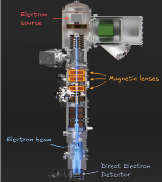
A cryo-EM column in cross-section: electrons fired from the source (top) are focused by magnetic lenses and captured by the Direct Electron Detector (bottom), tracing atomic shadows no light could ever cast. Courtesy: Branch Education (YouTube)
To capture these subatomic shadows, scientists developed the Direct Electron Detector—a camera where each microscopic pixel monitors a physical space smaller than a single atom. It snaps hundreds of frames per second, counting individual electrons one by one as they arrive. Supercomputers then strip away blur frame-by-frame, revealing a flawless, high-definition blueprint of the molecule.
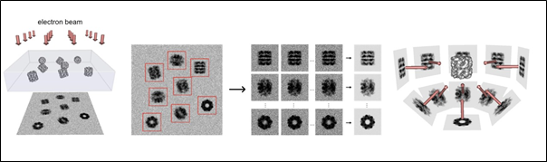
Various stages of Cryo-EM structure resolution. Courtesy: Greg Pintilie, CSAIL, MIT
Think of it like a smartphone sensor, but each microscopic pixel monitors a physical space smaller than a single atom. To prevent the vibrating protein from blurring, this camera operates at a blistering speed, snapping hundreds of frames per second to shoot a high-speed movie of the nanoscale world. It is so unfathomably precise that it literally counts individual electrons one by one as they strike the grid. By stripping away the blur frame-by-frame, supercomputers are left with a flawless, high-definition blueprint of the molecule.
Imaging moving molecules
But firing electrons at the protein is only half the battle. These biomolecules are suspended in water, constantly twisting and shifting. Photograph them at room temperature, and all you get is blur.
So, we, the structural biologists, freeze them. But freeze water normally, and it forms ice crystals—which at the microscopic level act like jagged knives, tearing delicate proteins apart.
To solve this, we plunge the fragile receptors into liquid ethane at nearly -196°C. They cool the sample so impossibly fast that the water molecules don’t have time to organize into crystals. Instead, the water instantly solidifies into a chaotic, glass-like state called vitreous ice.
This process quite literally stops time at the molecular level—capturing the shape-shifting proteins exactly as they were in that specific millisecond. Some resting, some open, some violently wedged. By firing the electron beam through this ice, then using supercomputers to stitch thousands of 2D images together, we can build a flawless 3D architecture of the receptor, atom by atom.
Building the Impossible Key
Structural biologists push this technology to near-atomic resolution for one very specific reason: precision medicine.
In the past, treating brain diseases was largely trial and error—flooding the body with chemicals and hoping something worked, often with devastating side effects. Today, we want to build a custom tool.
If we have an atomic map of a wedged receptor, we can design a drug molecule that acts as a microscopic corrective wedge—docking perfectly into an invisible crevice right next to the jammed amino acid, forcing the receptor to close smoothly again. But that docking site might exist for only a millisecond. Without knowing the exact spatial coordinates of every atom, the drug will simply bounce off.
This isn’t theoretical; it is already changing lives. By mapping the exact, high-resolution blueprint of a glutamate receptor (specifically the AMPA receptor), scientists discovered a hidden pocket on the protein’s surface. They then designed a breakthrough epilepsy drug called Perampanel—which wedges perfectly into that crevice, forcing the hyperactive receptor to close and halting devastating seizures.
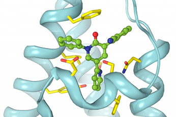
Perampanel (green), an anti-epileptic drug, wedged inside AMPA type glutamate receptor. Courtesy: CUIMC, Colombia University, USA
We cannot build precision medicine without atomic precision. By pushing the absolute limits of physics and microscopy, we are finally moving past the illusion that reading our DNA is enough. We are photographing the actual physical machinery of life, atom by atom—so we can finally learn how to repair it.